blog tambre
Karyotype analysis in assisted reproduction treatments

Table of contents
If you are undergoing assisted reproduction treatment or you are about to take the step of starting this wonderful adventure, you will surely have heard of karyotyping. The analysis of the karyotype is often crucial to make a good diagnosis and to find the keys to not achieving pregnancy, and that is why today we want to provide you with as much information as possible about it.
What do we call karyotype?
The karyotype is the complete set of chromosomes that an individual possesses. A “normal” karyotype is made up of 23 pairs of chromosomes, the last pair corresponding to the so-called ‘sex chromosomes’ (XY in the case of a male karyotype and XX in the case of a female karyotype). Health professionals recommend this test in order to detect abnormalities in the number of chromosomes or in their structure.
These anomalies can cause reproductive difficulties such as implantation failure or repeated miscarriages and it is therefore important to study them.
For whom is karyotyping indicated and how is it performed?
As mentioned above, there are chromosomal abnormalities that can lead to implantation failure or repeated miscarriages, as fertilisation and embryo development are affected. At Tambre we recommend that all women and couples undergoing In Vitro Fertilisation treatment should have a karyotype analysis, bearing in mind that they have already experienced complications in achieving a natural pregnancy or even in previous treatments.
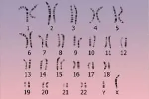
The karyotype test is extremely simple for patients, as all that is required is a blood test. The laboratory will then process the sample and create what is known as a karyogram or idiogram, which the doctor will interpret for the patient.
The above karyogram represents a karyotype in which there are no abnormalities, as we see 46 chromosomes showing no changes in their information. Next, we will explain the classification of other findings that we might find in a karyotype.
Chromosomal alterations in the karyotype
The alterations in a karyotype can be, as we explained at the beginning, related to the number of chromosomes or they can have to do with the structure of the chromosomes. Numerical anomalies occur when there is a greater or lesser number of chromosomes, as in the case of trisomies (presence of an extra copy of a specific chromosome, as occurs in Down’s syndrome with chromosome 21), monosomies (lack of a copy of a chromosome, as occurs in Turner’s syndrome with the absence of an X chromosome) or triploidies (existence of 69 chromosomes, which, in general, is incompatible with life).
Structural abnormalities sensitive to the karyotype analysis technique must be larger than 5 megabases (Mb). Smaller abnormalities can be detected by other tests, such as high-resolution karyotyping. The most common structural abnormalities that can be seen on a karyotype are as follows:
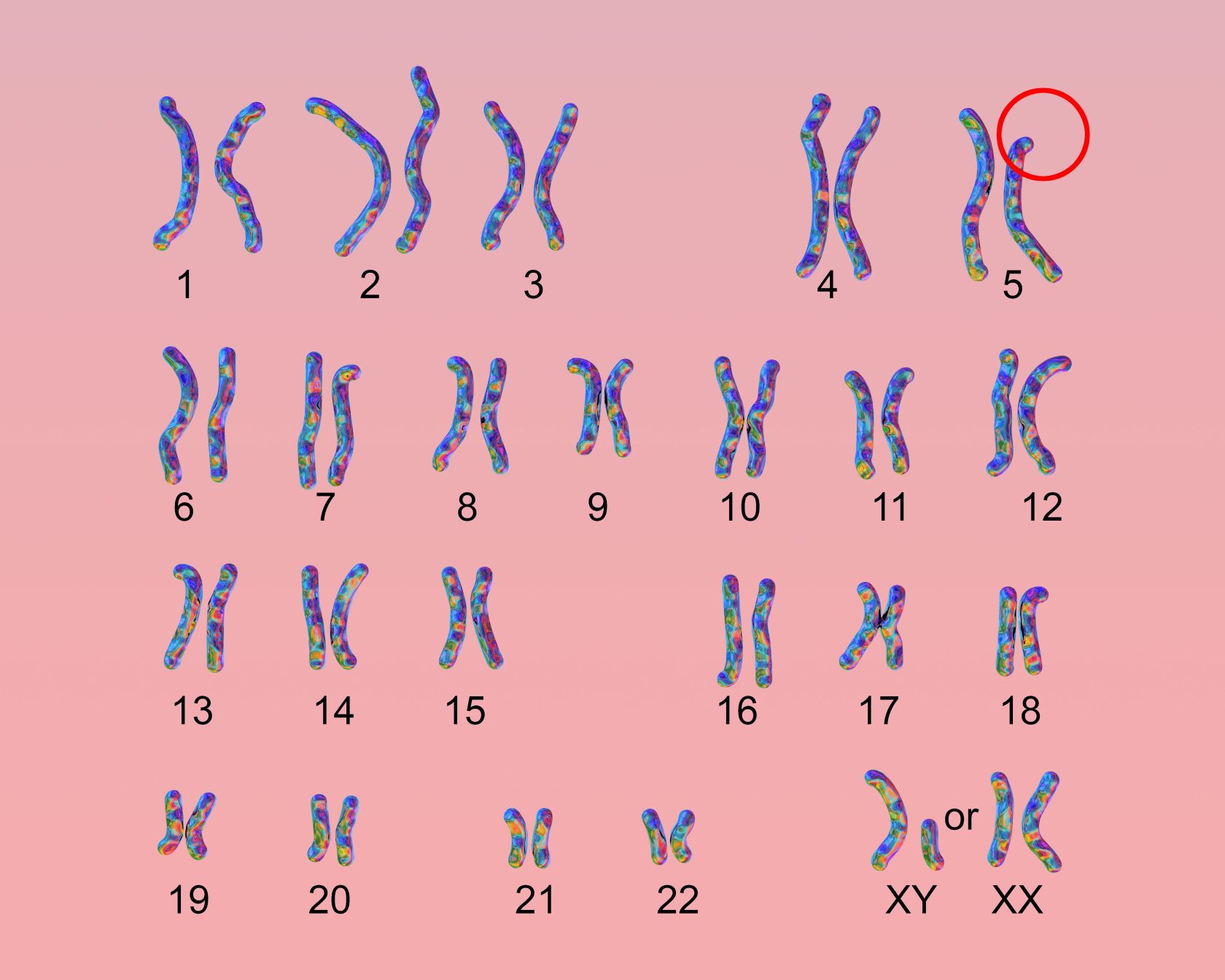
-Deletions: loss of DNA fragment material in a chromosome. Some of the most common deletions are those of chromosome 15, which are related to Prader-Willi Syndrome and Angelman Syndrome (depending on whether the origin is in the paternal or maternal chromosome respectively) and that of chromosome 5, which gives rise to Cat’s Meow Syndrome or cri du chat, whose karyogram can be seen below.
-Duplications: gain of DNA fragments on a chromosome (e.g. Pallister-Killian syndrome occurs if part of chromosome 12 is duplicated).
-Translocations: occur when a fragment of one chromosome is attached to another chromosome. Translocations are reciprocal when two different chromosomes swap fragments between them and are Robertsonian if one complete chromosome is linked to another.
-Inversions: occur when a chromosome segment changes its orientation within the chromosome itself.
If the karyotype studied has any of the anomalies explained throughout this article, assisted reproduction professionals will opt for the use of Preimplantation Genetic Testing techniques in the IVF cycle. If the alterations are more serious, the option of resorting to the clinic’s egg and/or sperm banks will be considered.
In short, karyotype analysis in assisted reproduction is a key test to achieve better results in fertility treatments and to detect possible congenital malformations or hereditary diseases in the offspring. If you have any doubts about this test or you think it could be useful in your case, do not hesitate to contact us!


